№ 21 · HEALTH
Dental X-rays: How Safe They Actually Are
June 06, 2026 · QDRO Team
Anyone who has ever sat in a dental chair with a film in their mouth and heard "hold still" has had a moment to wonder: how safe is this, really? X-rays are ionizing radiation, and that sounds alarming. But the doses involved are so small that they are hard to place alongside genuine health risks.
Let's compare the numbers. A bitewing — the standard radiograph that shows the crowns of the posterior teeth on both sides — delivers an effective dose of about 5 μSv (microsieverts). For reference: the natural radiation background in central Russia runs at roughly 7–8 μSv per day. A Moscow–New York flight adds about 70 μSv per trip. A single dental image is literally less than what you receive from the world around you over one day spent in the mountains.
But it is important to understand not only that the doses are small. It is important to understand why they are small, where in dentistry they are nonetheless higher, and what reasonable precautions exist.
How a safe dose is calculated, and what a microsievert is
The sievert is the unit of effective dose, and it accounts not only for the amount of energy absorbed but for the biological hazard that a particular type of radiation poses to particular organs. It is a biological measure of risk, not a physical one.
The International Commission on Radiological Protection (ICRP) has set an upper limit for the general public of 1 mSv (1,000 μSv) per year above natural background. The average annual dose from all medical procedures for a typical resident of a developed country is about 0.6 mSv. Dental radiography contributes a negligible share of it.
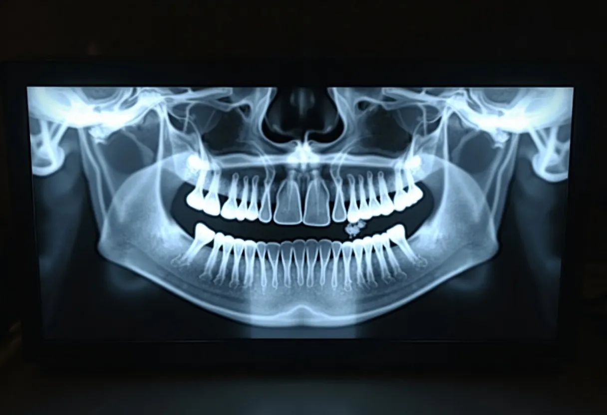
A full radiographic survey of the mouth — the orthopantomogram (a panoramic image of the entire dental arch) — delivers on the order of 14–24 μSv, depending on the equipment and the technical settings. The dosimetric study by Ludlow and colleagues, published in the Journal of the American Dental Association in 2008 (PMID 18762634), measured doses from the main types of dental radiography under the 2007 ICRP recalculation and showed that even a full set of periapical images comes to about 35 μSv with rectangular collimation and about 170 μSv with round collimation — still far below the annual limit for the public.
Measurement of effective doses from the main types of dental radiography on a phantom. A full set of periapical images with round collimation (~170 μSv) remains far below the annual limit for the public of 1,000 μSv; switching to rectangular collimation lowers the dose to roughly 35 μSv.
CBCT: a different conversation
Cone beam computed tomography (CBCT) is a separate story. The method produces a high-resolution three-dimensional image of the teeth, roots, alveolar bone, and joints. It is used for implant planning, orthodontic diagnosis, and the assessment of impacted wisdom teeth.
The doses here are substantially higher. According to the dosimetric study by Pauwels and colleagues within the SEDENTEXCT project (European Journal of Radiology, 2012, PMID 21196094), which measured doses on 14 CBCT units, the effective dose ranges from 19 to 368 μSv depending on the field of view (FOV), the exposure protocol, and the specific machine. Units with a small field of view (small FOV, covering one or two teeth) deliver considerably less radiation than units with a large field that captures the whole skull.
The ALARA principle ("As Low As Reasonably Achievable") means exactly this in radiology: the minimum diagnostically sufficient dose, not zero dose at any cost.
The thyroid gland: why the collar goes on
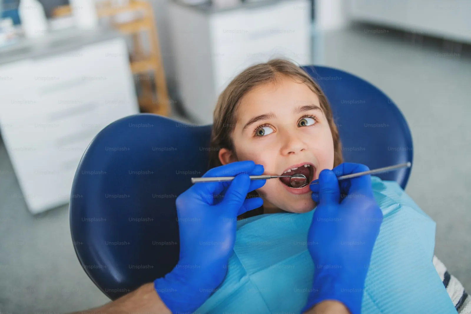
The thyroid is one of the most radiosensitive organs, especially in children. During dental radiography it sits close to the primary beam, though not in its main path. A lead thyroid collar shields it from scattered and direct radiation.
The study by Han and colleagues (PMID 24005060, Dentomaxillofacial Radiology, 2013) measured the thyroid dose during digital panoramic radiography on four machines. Without shielding, the thyroid receives on the order of 1–3 μSv — negligibly little, but technically measurable. A lead collar placed at the front of the neck lowers that dose further.
The American Dental Association (ADA) and the American Academy of Oral and Maxillofacial Radiology (AAOMR), in their joint recommendations on patient selection for dental radiography (Benavides E et al., Oral Surg Oral Med Oral Pathol Oral Radiol, 2026, PMID 41581943), state that shielding and justified patient selection are the standard of care, particularly for children, pregnant patients, and people with heightened radiosensitivity.
How often images can be taken
There is no universal answer, and that matters fundamentally. Dental associations explicitly warn against "routine annual radiographs" without clinical justification.
The ADA and AAOMR, in their patient selection recommendations (PMID 41581943), divide patients into risk groups:
- Children with no erupting permanent teeth and low caries risk: bitewing images every 1.5–2 years.
- Asymptomatic adults at low risk: a full survey image every 2–3 years; bitewings every 2 years are sufficient.
- High caries risk (active decay, xerostomia, immunodeficiency): imaging more often — bitewings every 6–12 months.
- A new patient at the practice with no recent images: a full survey image for baseline diagnosis is justified regardless of risk category.
The key phrase is "clinical justification." If a patient has no complaints, no visible lesions, and no risk factors, taking a panoramic image every year as a box-ticking exercise is unnecessary from both a clinical and a radiological standpoint.
The cumulative picture looks like this: even under the most intensive monitoring (four bitewing images every six months plus an annual panoramic image), the yearly dose from dental radiography comes to roughly 80–100 μSv — that is, about 10% of the permissible annual limit for the public, and several times less than a year of background dose.
Pregnancy: real risk or mythology
Pregnancy is a special case. The fetus is considerably more sensitive to ionizing radiation than an adult, and that vulnerability is at its highest in the first trimester.
Here too, the numbers put everything in its place. The ADA/AAOMR patient selection recommendations (PMID 41581943) confirm that with a standard lead apron the dose to the uterus and fetus from a bitewing image is practically indistinguishable from zero — less than 0.01 μGy. That is hundreds of thousands of times below the threshold at which teratogenic effects arise in a fetus (thought to lie in the region of 100 mGy, that is, 100,000 μGy).
If a pregnant woman has a toothache, leaving her untreated is a more serious risk than taking one periapical image with shielding. The stress response to pain and infectious processes in the mouth pose a considerably greater threat to the fetus than the radiation dose from a single image.
This is not a call to recklessness — it is a call to weigh risks rationally. Elective X-ray images are better postponed until after delivery. Urgent ones should be taken with shielding, without hesitation.
Childhood: why it matters not to overdo the protection
The paradox of radiation protection is that excessive fear of X-rays can do more harm than the images. Undetected decay in a child, missed because the parents refused an X-ray, is a real deterioration in the health of the teeth and the bite.
Children do receive a somewhat higher effective dose than adults from the same image, because of their smaller body size and the higher radiosensitivity of a number of tissues. But here too the absolute values remain small.
The key measure is using a rectangular collimator instead of a round one. This technical step (it limits the beam to the size of the film or sensor) reduces the dose by 60–70% with no loss of diagnostic value. PMID 18762634 records this directly: a full set of images with round collimation delivers ~170 μSv, and with rectangular collimation about 35 μSv — which makes switching to a rectangular collimator one of the most effective technical measures for lowering dose in dental radiography.
Digital radiography instead of film is the second most significant dose-reducing factor, and it is available in most clinics today.
To sum up: dental radiography in its standard forms is one of the least hazardous sources of ionizing radiation in medicine. The sensible approach is not to refuse it, but to take images on indication, use shielding, choose digital equipment, and avoid confusing "ionizing" with "dangerous" at the doses in question.
At QDRO we believe that informed dental care begins with an understanding of the real picture — not with anxiety built on misread facts.
Sources:
- PMID 18762634 — Ludlow JB et al., J Am Dent Assoc, 2008 — measurement of effective doses from the main types of dental radiography under the 2007 ICRP recalculation, including the effect of collimation
- PMID 25270063 — Horner K et al., Dentomaxillofac Radiol, 2015 — guidelines on the clinical use of CBCT, summarizing the principles of the SEDENTEXCT project
- PMID 41581943 — Benavides E et al., Oral Surg Oral Med Oral Pathol Oral Radiol, 2026 — ADA/AAOMR recommendations on patient selection for dental radiography and CBCT, including pregnant patients and children
- PMID 24005060 — Han GS et al., Dentomaxillofac Radiol, 2013 — shielding effect of the thyroid collar in digital panoramic radiography
- PMID 21196094 — Pauwels R et al., Eur J Radiol, 2012 — range of effective doses in cone beam computed tomography (SEDENTEXCT project, 14 units)